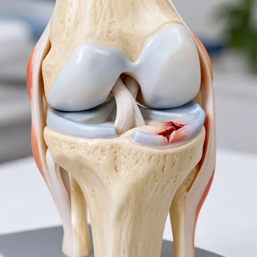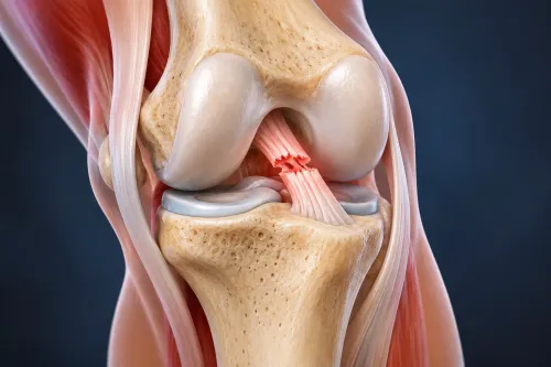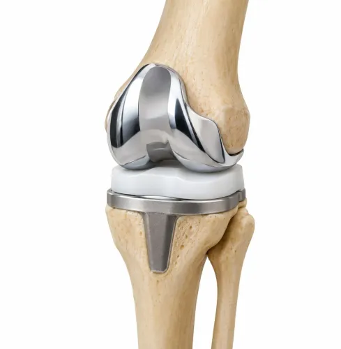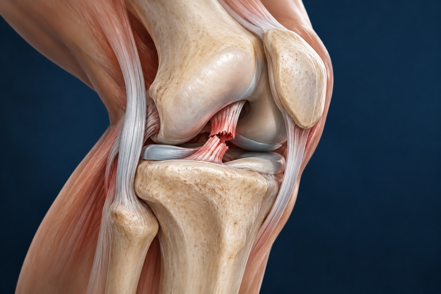
A knee that gives way after injury can reflect damage to one ligament, several ligaments, or structures such as the meniscus and cartilage. By the end, you will know how instability is confirmed, when rehabilitation or bracing may be enough, how reconstruction is performed, and which personal factors shape the decision.
Key takeaways
- Confirm instability with repeated giving-way episodes and abnormal ligament tests.
- Use MRI to assess ligament damage and X-rays to check bone alignment.
- Treat swelling and restore knee movement before reconstruction when possible.
- Expect staged rehabilitation focused on motion, strength, balance and return to activity.
Which injuries cause a knee to give way, and how is instability confirmed?
Clinicians confirm true knee instability by matching repeated giving-way episodes with abnormal ligament tests, not by pain alone. A swollen knee can inhibit quadriceps control and buckle during walking, while a stiff knee can feel unstable because it cannot fully bend or straighten.
The history records the injury mechanism, direction of force, immediate swelling, locking, and whether episodes continue after swelling settles.
- Lachman test: forward movement of the tibia compared with the femur points to an ACL injury.
- Pivot-shift test: a slipping or clunking sensation during rotation supports functional ACL insufficiency.
- Posterior drawer test: backward tibial movement indicates a PCL tear.
- Valgus stress test: opening on the inner side suggests an MCL tear.
- Varus stress test: opening on the outer side suggests an LCL tear.
- Range-of-motion testing and quadriceps strength help separate mechanical laxity from stiffness or weakness.
- Comparing each result with the uninjured knee helps identify pre-existing looseness.
| Injury | Examination pattern | Interpretation |
|---|---|---|
| Isolated ACL tear | Positive Lachman or pivot shift | Anterior or rotational instability |
| PCL tear | Positive posterior drawer | Posterior sag or backward instability |
| MCL tear | Valgus opening | Medial instability |
| LCL tear | Varus opening | Lateral instability |
| Combined ligament injury | Abnormal results in several directions | Greater instability requiring specialist assessment |
Painful guarding can obscure these findings, so clinicians may repeat the examination after swelling and pain decrease. A single buckle during severe pain does not prove ligament failure; recurrent, direction-specific giving way with laxity does.
Which scans are useful, and when is surgery the right treatment?
MRI knee imaging shows whether the ACL, PCL, MCL, or LCL is torn and identifies meniscal, cartilage, bone-bruising, and associated injuries. It does not measure how often the knee gives way.
Standing X-rays reveal fractures, joint-space loss, alignment, and arthritis under load; stress radiographs apply force while images record ligament laxity compared with the other knee.
A CT scan knee instability assessment is most useful for bone detail, tunnel position after previous surgery, fractures, rotational alignment, or complex multi-ligament injury. CT does not replace MRI for menisci or ligament fibres.
| Option | What it involves | When it applies |
|---|---|---|
| Structured rehabilitation | Strength, range of motion, balance, and movement training | Isolated tear with manageable symptoms and no repeated giving way |
| Bracing | External support during healing or activity | Selected collateral-ligament injuries or temporary protection |
| Repair | Reattaching a repairable ligament near its attachment | Certain fresh tears with good tissue quality |
| Reconstruction | Replacing a failed ligament with a graft | Persistent instability, high-demand activity, or combined injury |
| Observation | Activity modification and review without an operation | Stable knee, low demands, or symptoms improving |
Surgery is not automatic after an ACL tear. Repeated giving-way, a repairable meniscal tear, combined ligament damage, or goals involving cutting sports can shift the balance toward reconstruction. Preserving a meniscus can slow early rehabilitation because weight bearing or knee flexion may be restricted.
Rehabilitation first can produce similar two-year outcomes to routine early reconstruction for many acute ACL injuries. Before surgery, swelling should settle and full extension return; operating on a stiff knee raises the risk of persistent motion loss. Reconstruction improves stability, but it does not guarantee protection from later arthritis or another tear.
Why timing and graft choice matter before reconstruction
A swollen knee with lost extension is a poor setting for reconstruction. Surgeons may first use rehabilitation, ice, compression, and guided exercises to reduce swelling and restore full straightening, because operating on a stiff knee raises the risk of arthrofibrosis and lasting motion loss.
Knee ligament reconstruction timing is not automatically “as soon as possible”: initial rehabilitation with optional delayed ACL reconstruction produced similar two-year outcomes to routine early surgery in many patients. Earlier surgery may still suit persistent giving-way, a repairable meniscal tear, or combined ligament damage.
The graft replaces the torn ACL; the usual operation does not stitch the original ligament together. Fixation hardware holds the graft while it incorporates and remodels, so a technically successful reconstruction does not make the knee immediately normal.
| Graft | Fixation and healing | Failure risk | Donor-site symptoms |
|---|---|---|---|
| Patellar tendon graft | Bone plugs allow strong bone-to-bone fixation | Low failure rates | More anterior-knee pain and difficulty kneeling |
| Hamstring autograft | Soft-tissue fixation requires tendon-to-bone healing | Different failure pattern from patellar tendon grafts | Less kneeling pain, but possible hamstring weakness |
| Quadriceps-tendon autograft | Strong tendon tissue; fixation depends on whether a bone block is used | Established option, with less long-term comparative evidence | Avoids previously harvested sites; front-thigh weakness or pain can occur |
| Allograft | Donor tissue avoids harvest, but biological incorporation can be slower | Higher failure risk in younger, active patients | No graft-harvest wound or related weakness |
For younger or highly active patients, an autograft is usually preferred over an allograft. The choice among autografts balances stability against pain, kneeling demands, muscle weakness, and previous harvest sites.
What happens during reconstruction, and what does recovery require?
An ACL reconstruction replaces the torn ligament with a tendon graft; it does not usually stitch the original ACL back together. During arthroscopic ligament reconstruction, the surgeon inspects the joint, creates tunnels through the tibia and femur, passes the graft into the correct anatomical position, and secures it with screws, buttons, or another fixation device.
1. The surgeon checks the meniscus and cartilage through the arthroscope. A repairable meniscus tear may receive sutures, while an irreparable flap may require limited trimming; cartilage defects may be stabilised or treated according to their size and location.
2. Graft fixation holds the tendon while it bonds to the bone and remodels. A stable operation does not make the knee immediately normal, and it does not eliminate later instability, meniscal damage, cartilage symptoms, or osteoarthritis risk.
3. Rehabilitation restores extension, bending, swelling control, quadriceps strength, balance, and controlled landing. Meniscus repair can require crutches, reduced weight bearing, or limits on knee flexion, making recovery slower than after isolated ACL reconstruction.
4. Before return to sport after ACL reconstruction, you need a quiet knee with full motion, stable ligament tests, near-symmetrical strength, and satisfactory single-leg hop, landing, agility, and change-of-direction performance. Your clinician also assesses movement quality and confidence under fatigue; time alone is not clearance for pivoting sports.
Recovery is measured in phases, not by pain alone. Returning before strength and movement control recover raises reinjury risk, so follow-up testing decides when running, cutting, contact, and competition can resume.
Which personal factors change the reconstruction plan and prognosis?
Age alone does not decide whether reconstruction is worthwhile. Ask how much pivoting, kneeling, climbing, lifting, or uneven-ground work your knee must tolerate, and whether you can complete rehabilitation. A desk worker, competitive footballer, and construction worker may need different stability targets.
| Factor | How it changes the plan | What to discuss |
|---|---|---|
| Age and activity | Graft choice, rehabilitation pace, and return-to-sport testing | Your sport, work demands, and realistic activity level |
| Growth plates | A growth plate-safe reconstruction protects remaining bone growth | Your skeletal maturity and risk of growth disturbance |
| Knee alignment | Bow-leg or knock-knee alignment can overload a reconstructed ligament | Whether standing X-rays show a need to address alignment |
| Smoking or diabetes | Nicotine impairs healing; poorly controlled glucose raises infection risk | A quit plan and your HbA1c before surgery |
| Previous surgery | Tunnel position, hardware, graft availability, and scar tissue complicate revision ligament reconstruction | Whether staged surgery or a different graft is needed |
| Meniscus or cartilage injury | Meniscal repair can restrict weight bearing or knee flexion and slow rehabilitation | Whether preserving the meniscus changes your schedule |
A technically successful graft does not make the knee immediately normal or prevent osteoarthritis. ACL reinjury prevention depends on strength, movement control, and passing functional tests; returning to pivoting sport before nine months carries greater risk in young-athlete studies, but nine months is not a personal guarantee.
Dr Ronak Kothari can help weigh these factors against your examination, scans, work demands, and willingness to complete rehabilitation rather than treating the scan alone.
Related service
Knee Arthroscopy & Sports Medicine Knee Arthroscopy & Sports Medicine Dr. Ronak Kothari, a skilled Orthopaedic Surgeon, specializes in Knee Arthroscopy and Sports Medicine, offering advanced, minimally invasive... View service → |
Frequently asked questions
Which injuries can cause the knee to give way?
ACL, PCL, MCL and LCL injuries can cause instability. Meniscus damage, swelling, weakness and restricted movement can also make the knee buckle.
How do clinicians confirm true knee instability?
They combine your history of repeated giving-way episodes with ligament tests such as the Lachman, pivot-shift, posterior drawer and varus or valgus stress tests.
Which scans are useful before knee ligament reconstruction?
X-rays show fractures, arthritis and alignment. MRI shows ligament tears, meniscus damage, cartilage injury and other soft-tissue problems.
When is surgery the right treatment for knee instability?
Reconstruction is considered when instability persists, the knee has a repairable injury pattern, and rehabilitation alone cannot support your work, sport or daily activities.
What affects recovery after ligament reconstruction?
Timing, graft choice, the injured ligaments, knee motion, muscle strength, age, general health, smoking and adherence to rehabilitation all affect recovery and prognosis.